
Chapter 55: Ischemia
Vasculitis
The vasculitides are a diverse group of diseases that cause a proportionately higher incidence of cerebral ischemic events in young and middle-aged adults than in the elderly. In one series, approximately 7% of strokes occurring in patients between the ages of 15 and 40 years were the result of vasculitis.54 Vasculitis is characterized by necrosis and inflammation of the vessel wall that can eventually result in segmental alteration of the vessel caliber, occlusion, aneurysm, and areas of hypervascularity.79
There are inherent difficulties in classifying the vasculitides because of their large overlap of clinical features and pathological characteristics. The vasculitis may be the primary disease process (e.g., polyarteritis nodosa) or may be a secondary manifestation of a generalized systemic disease (e.g., systemic lupus erythematosus). However, for the purposes of this chapter, we place the vasculitides into two major categories—infectious and noninfectious.
Infectious cerebral vasculitis can occur as a result of direct spread from initial meningeal or parenchymal infection or secondary to hematogenous seeding. Many infections, including purulent bacterial meningitis, tuberculosis, and syphilis, as well as those infections caused by viral agents (e.g., herpes) and an increasing number of fungal agents (e.g., Cryptococcus and Coccidioides organisms) as a result of the relatively large number of immunocompromised patients, result in cerebral arteritis (see Chapter 60).
The noninfectious vasculitides can be further characterized based on the immunological mechanism of the disease process and divided into three general categories—those caused by immune complex deposition, those caused by cell-mediated mechanisms, and those associated with diverse syndromes (Table 55-1).78 It is beyond the scope of this chapter to fully discuss the various noninfectious vasculitides, but a brief discussion of systemic lupus erythematosus (SLE) and polyarteritis nodosum (PAN) and their MR appearances follows the general discussion.
| Probable immune complex deposition mechanism |
|---|
| Systemic necrotizing vasculitis |
| Polyarteritis nodosa |
| Allergic angiitis and granulomatosis |
| Overlap syndrome |
| Hypersensitivity vasculitis |
| Serum sickness or serum sickness–like vasculitis |
| Henoch-Schönlein purpura |
| Collagen vascular disease with vasculitis |
| Hypocomplementemic vasculitis |
| Malignancy with vasculitis |
| Mixed cryoglobulinemia |
| Probable cell-mediated mechanism with round cell granuloma formation |
| Giant cell arteritis |
| Temporal arteritis |
| Takayasu's arteritis |
| Wegener's granulomatosis |
| Lymphomatoid granulomatosis |
| Miscellaneous |
| Mucocutaneous lymph node syndrome (Kawasaki syndrome) |
| Thromboangiitis obliterans (Buerger's disease) |
| Behçet's disease |
| Cogan's syndrome |
| Sweet's syndrome |
| Erythema nodosum |
| Bowel bypass dermatitis |
| From reference 78. |
The angiographic appearance of vasculitis is often nonspecific and can be mimicked by such varied entities as atherosclerosis, vasospasm as a result of subarachnoid hemorrhage, and neoplastic vascular involvement (e.g., gliomas and lymphomas).45 In a recent study by Harris and co-workers57 comparing brain MRI and invasive cerebral angiography in diagnosing cerebral vasculitis, the MR examination was abnormal in all the patients diagnosed with vasculitis who had both studies. Yet, the conventional angiogram has a false-negative rate of 27% and a diagnostic yield of only 6%. The authors concluded that a negative MRI was able to exclude intracranial vasculitis more definitively than a negative angiogram and furthermore makes the likelihood of finding vasculitis with angiography alone negligible.
The most common finding seen on MRI in the nonseptic vasculitides is generalized cerebral atrophy.72 However, the nonseptic vasculitides encompass a broad spectrum of findings on MRI because a given disease process may involve arteries, veins, or both and often affects vessels of a particular size and location, thereby displaying a distribution of pathology, which though nonspecific is suggestive of a diagnosis.48,74,115 Small vessel ischemic disease is the most common lesion that may mimic the nonseptic vasculitides. Small vessel ischemic disease is seen in the elderly population, particularly in those who are hypertensive. The ischemic lesions have a predilection for the centrum semiovale, basal ganglia, and pons.59 The subcortical U-fibers tend to be spared because they are supplied by intermediate-length arterials and are thus less susceptible to the effects of hypotension. In contrast, cerebral vasculitis tends to occur in young adults, can involve the subcortical white matter, and can be hemorrhagic.50
Systemic Lupus Erythematosus.
SLE is a disease of unknown origin characterized by a plethora of autoantibodies to cellular proteins and nucleic acids. SLE predominately affects women (female/male ratio of 9:1) and usually arises in the second and third decades of life but may become manifest at any age.104 Involvement of the CNS is a common manifestation of SLE, occurring in 50% to 70% of lupus patients.13 Symptoms include migraine headaches, seizures, and psychosis, as well as focal neurological signs. Approximately 15% of lupus patients suffer stroke, usually within the first few years of diagnosis.43,87 However, true CNS lupus vasculitis is rare.13 Rather the most common finding seen at CNS autopsy is perivascular inflammation of small vessels or endothelial cell proliferation.13 Most strokes in lupus patients are probably caused by emboli either from their verrucous endocarditis, or hypercoagulability as a result of antiphospholipid and anticardiolipin antibodies.59
On MRI the most common finding is mild to moderate atrophy seen in 67% of SLE patients. In addition, three patterns of focal involvement have been identified: (1) large cerebral infarctions involving gray and white matter in the territory of a major vessel, usually the MCA; (2) small white matter infarctions, which are often multiple in a periependymal distribution; and (3) small cortical gray matter lesions, which often resolve in 2 to 3 weeks (Figure 55-26). Whether the small gray matter lesions represent reversible ischemia or a temporary alteration in vascular permeability has not been determined.1 Subarachnoid hemorrhage, cerebral vein thrombosis, aseptic and septic meningitis, and cerebral abscess also occur in lupus patients with some frequency.1,76,125
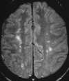 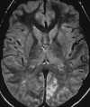 |
| Figure 55-26 A 29-year-old woman with SLE. A, Proton density–weighted axial image shows patchy punctate foci of hyperintensity involving the subcortical white matter. B, More focal abnormality exists in the calcarine cortex in this patient with left visual field hemianopsia. |
Polyarteritis Nodosa.
Polyarteritis nodosa (PAN) is an idiopathic, necrotizing inflammatory disease that may affect any small or medium-sized artery in any organ system of the body. The characteristic saccular microaneurysms seen in PAN are the result of internal elastic lamina necrosis, yet this diagnostic hallmark is seen in only 60% to 75% of patients with the disease.36 CNS involvement is typically a latent manifestation and has been reported in 8% to 44% of patients with PAN.74 Because of the multiple vessel involvement of PAN, the MR findings are nonspecific and consist of both major and nonmajor vascular territory ischemia/infarction.74
Sickle-Cell Disease
Cerebrovascular complications have been reported to be as high as 26% in patients with sickle-cell disease (SCD). The principal cerebrovascular manifestations are cerebral infarction (75%), intracerebral hemorrhage (20%), and subarachnoid hemorrhage (1% to 2%).98 The pathogenesis of cerebrovascular disease in SCD remains incompletely understood. Initially, cerebral infarction in SCD was thought to be caused by occlusion of small vessels by sludging of sickled red blood cells. More recent research proposes that endothelial injury from the abnormal adherence of sickled erythrocytes to the endothelium is the initiating event in arterial wall injury.98 Subsequently there are internal elastic lamina fragmentation and degeneration of the muscularis, which ultimately result in large vessel vasculopathy and aneurysm formation.98 At autopsy, approximately 50% of SCD patients with infarction show thrombi, primarily involving the distal cervical and proximal internal carotid arteries.106 Infarcts predominate in the distal branches of the ICA (ACA and MCA territory) and at the ACA-MCA watershed zone. It is thought that the predilection for the ACA-MCA watershed zone is the result of hemodynamic insufficiency superimposed on large vessel vasculopathy. Patients with significant endarteritis and stenosis of the proximal cerebral vessels are at significant risk (reported to be as high as 67%) for recurrent stroke.98,136 These patients are at higher risk for complications during conventional cerebral angiography and need to be well hydrated, transfused, and often sedated before the procedure. MRA therefore can be quite helpful in the depiction of large vessel vascular occlusion or stenosis in the acute setting. Thus both MRI and MRA are helpful in the early detection of cerebrovascular complication, enabling prophylactic transfusion therapy to be initiated in an attempt to mitigate an otherwise high recurrence rate and poor prognosis.98,136
Moyamoya Disease
Moyamoya disease, first described in Japanese children, has since been diagnosed in all races and in adults. Anatomically this disease is characterized by stenosis or occlusion of the distal ICA, the proximal MCA and ACA, and less commonly the proximal PCA. This occlusion results in a network of dilated collateral vessels, usually lenticulostriate and thalamoperforating arteries, that on angiography have a vascular blush resembling a puff of smoke, or moyamoya in Japanese (Figure 55-27). Pathologically this occlusion of the large arteries around the circle of Willis is secondary to a noninflammatory intimal hyperplasia, fibrosis, and thrombosis. However, the cause of the disease remains unknown. A moyamoya pattern of collateral blood flow is nonspecific because it can be seen in other slowly progressive intracranial vascular occlusive entities, such as atherosclerosis, neurofibromatosis, and sickle-cell disease. The disease is progressive, and the clinical course is ultimately determined by the effectiveness of the collateral circulation.
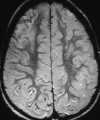 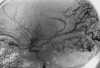 |
| Figure 55-27 Moyamoya disease. A, Proton density– weighted axial image shows gyral hyperintensity involving the right frontal lobe and, to a lesser extent, the left frontal lobe. Subtle signal alteration is also seen involving the white matter. B, Angiogram documents occlusive disease of the right ICA with proliferation of collateral perforators typical of moyamoya disease. |
The most common cerebral lesions demonstrated by MRI in patients with moyamoya disease are multiple, often bilateral, cortical and subcortical watershed distribution infarctions in the ICA territory. In addition, deep white matter lesions, which are often multiple, bilateral, and asymptomatic, occur in the majority of patients. MRA is useful because it can depict the major collateral flow patterns, as well as most large vessel occlusions.132 Enlargement of the small collateral vessels, particularly in the basal ganglia, can be demonstrated on T1-weighted axial images as numerous small circular or serpentine foci of signal void.26,27
Summary
From this discussion it is clear that MRI has advanced beyond the stage of simply depicting neuroanatomy and is also capable of characterizing the dynamic pathophysiological processes associated with ischemia. Significant research efforts now focus on the MR identification of salvageable, at-risk brain parenchyma. Combined MRI and spectroscopy should offer important diagnostic and prognostic information in the acute stroke patient. In addition, these combined methods will likely have a key role in the development and assessment of various stroke therapies, which could ultimately result in a significantly improved patient outcome.
~ Previous ~ Next ~
~ Back to Chapter Index ~
~ Back to Magnetic Resonance Imaging main page ~